ABSTRACT
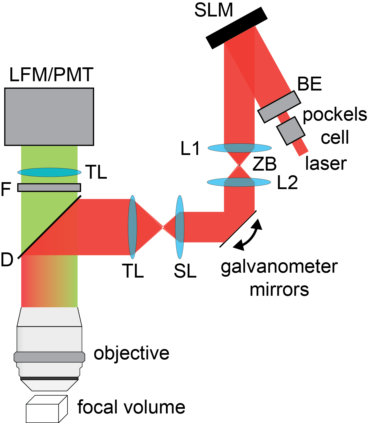
Phase spatial light modulators (SLMs) are widely used for generating multifocal three-dimensional (3D) illumination patterns, but these are limited to a field of view constrained by the pixel count or size of the SLM. Further, with two-photon SLM-based excitation, increasing the number of focal spots penalizes the total signal linearly—requiring more laser power than is available or can be tolerated by the sample. Here we analyze and demonstrate a method of using galvanometer mirrors to time- sequentially reposition multiple 3D holograms, both extending the field of view and increasing the total time-averaged two-photon signal. We apply our approach to 3D two-photon in vivo neuronal calcium imaging.
FILES
CITATION
Samuel J. Yang, William E. Allen, Isaac Kauvar, Aaron S. Andalman, Noah P. Young, Christina K. Kim, James H. Marshel, Gordon Wetzstein, and Karl Deisseroth, “Extended field-of-view and increased-signal 3D holographic illumination with time-division multiplexing,” Opt. Express 23, 32573-32581 (2015)
BibTeX
@article{Yang:15,
author = {Samuel J. Yang and William E. Allen and Isaac Kauvar and Aaron S. Andalman and Noah P. Young and Christina K. Kim and James H. Marshel and Gordon Wetzstein and Karl Deisseroth},
journal = {OSA Optics Express},
number = {25},
pages = {32573–32581},
title = {Extended field-of-view and increased-signal 3D holographic illumination with time-division multiplexing},
volume = {23},
year = {2015},
}